
Polyorchidism: sonographic and magnetic resonance imaging findings
DOI :
https://doi.org/10.5489/cuaj.704Résumé
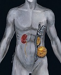
Polyorchidism is a rare anomaly and frequently associated withcriptorchidism, inguinal hernia and testicular torsion. It is also
reported as increased risk of testicular malignancy. We report a
case of 23 year old man with left supernumerary testis in the left
hemiscrotum. He presented with painless mass in his left hemiscrotum.
Normal physical examination and laboratory tests including
spermiogram were examined. Both ultrasound and MRI examinations
revealed polyorchidism without malignancy or any other
concomitant features. In most cases sonography alone is diagnostic.
MRI may provide additional information in complicated cases of
polyorchidism. Conservative treatment with sonographic follow-up
is the choice of treatment in uncomplicated cases.
Téléchargements
Les données relatives au téléchargement ne sont pas encore disponibles.
Téléchargements
Comment citer
Yalçınkaya, S., Şahin, C., & Şahin, A. F. (2013). Polyorchidism: sonographic and magnetic resonance imaging findings. Canadian Urological Association Journal, 5(5), E84-E86. https://doi.org/10.5489/cuaj.704
Numéro
Rubrique
Case Report
Licence
Les auteurs accordent les droits d’auteurs liés à l’article et son contenu à l’Association des urologues du Canada. Cette entente signifie que vous ne pouvez pas faire ce qui suit, sans d’abord obtenir l’autorisation écrite de l’AUC :
- Afficher l’article sur tout site Web.
- Traduire ou autoriser une tierce partie à traduire l’article.
- Copier ou reproduire l’article par quelque moyen que ce soit et sous tout format que ce soit, ou autoriser d’autres à le faire, au-delà de ce qui est permis par la loi canadienne du droit d’auteur.
- Copier ou reproduire des sections de l’article, y compris les tableaux et figures, par quelque moyen que ce soit, ou autoriser d’autres à le faire, au-delà de ce qui est permis par la loi canadienne du droit d’auteur.
L’AUC encourage l’usage des articles à des fins éducatives sans but commercial et ne refusera pas sans motif raisonnable toute demande d’autorisation à cet effet.
Vous conservez le droit moral lié à l’article et son contenu. Cela signifie que l’AUC ne peut utiliser ses droits d’auteurs d’une manière telle que cela pourrait avoir des répercussions négatives sur votre réputation ou sur votre droit à être associé à l’article.
L’AUC exige également que vous garantissiez ce qui suit :
- Vous êtes l’auteur ou les auteurs et seul(s) propriétaire(s) du contenu, le contenu de l’article est original et n’a jamais été publié et vous n’en avez pas déjà cédé les droits d’auteurs ni accordé de licence concernant son contenu à toute autre tierce partie;
- Toutes les personnes qui ont contribué de manière considérable à la rédaction de l’article sont mentionnées;
- L’article ne viole aucun droit de propriété de toute tierce partie, et vous avez obtenu les autorisations requises pour inclure les travaux d’autres personnes dans cet article; et
- L’article ne diffame aucune tierce partie ni ne viole les droits à la vie privée de toute tierce partie.
